EAR INFECTION:
- 1.Otitis Externa (Swimmer’s ear)
- 2.Otitis Media
- a) Acute Otitis Media
- b) Chronic Otitis Media
- 3.Labyrinthitis
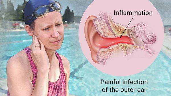
1.OTITIS EXTERNA:
^Cause inflammation (redness and swelling) of the external ear canal(EAC), which is the tube between the outer ear and eardrum.
^Called as swimmer’s ear as repeated exposure to water can make ear canal more vulnerable to inflammation.

Symptoms:
- ^Ear pain, can be severe. (Otalgia)
- ^itchiness in the ear canal (Pruritus)
- ^a discharge of liquid or pus from the ear (Otorrhea)
- ^some degree of temporary hearing loss
- ^tenderness to palpation
- ^usually one ear is affected
Causes:
- ^bacterial infection (most common) (Pseudomonas aeruginosa and Staphylococcus aureus).
- ^irritation.
- ^fungal infections (Otomycosis) (Aspergillus, Candida).
- ^allergies.
- ^damaging skin inside your ear (due to cotton wool buds)
- ^regularly getting water in your ear (ideal environment for bacteria to grow).
Prevention:
^Instruct Patient to not use cotton swabs or any other objects to canal.
^Swimmers are to be instructed to use ear plugs and advised to use alcohol-vinegar (1:1) drops after swimming.
Ix:
^Otoscopy (reveals mycelia establishing diagnosis of Otomycosis)
^Lab: typically not needed, gram staining and culture of auditory canal can help in patients with immunocompromised status.
Cx:
- ^Abscess
- ^Stenosis of ear canal (due to thick and dry skin build inside ear canal due to chronic OE)
- ^Inflamed or perforated eardrum (spread of infection to ear drum causing tear and symptoms such as: temporary hearing loss, earache or discomfort, a discharge of mucus from ear, ringing or buzzing in your:tinnitus)
- ^Malignant otitis external, infection spreads from the ear canal into the surrounding bone, requires prompt treatment with antibiotics and sometimes surgery, can be fatal if left untreated.
Rx:
- ^sometimes can self-resolve, but takes several weeks
- ^NSAID, Opioids or topical steroid preparations (for pain)
- ^Antibiotic drops (Ofloxacin, Ciprofloxacin, Colistin, Polymyxin B, Neomycin, Chloramphenicol, Gentamicin and Tobramycin.)
- ^Polymyxin B and Neomycin preparations are often used in combination for the treatment of S Aureus and P Aeroginosa Infections.
- ^Steroid ear drops helps reduce edema and otalgia
- ^Otomycosis Rx includes cleansing and debriding the EAC, acidifying the canal, and administering anti fungal agents.
- ^Non specific Antifungal (Merthiolate)
- ^Specific anti fungal (clotrimazole, Nystatin, Ketoconazole)
- ^Itraconazole is orally administered, effective against Aspergillus.
- ^Aural packing (Ear wick placement) and Antibiotic/combination preparation application – 4 times a day like 3-4 drops, changed every day.
Treatment Guidelines:
- Acute otitis externa should be distinguished from other possible causes of ear canal inflammation.
- Topical antimicrobial otic preparations should be considered the first-line treatment for uncomplicated acute otitis externa.
- Addition of a topical corticosteroid may result in faster resolution of symptoms such as pain, canal edema, and canal erythema.
- Systemic antibiotics should be used only if the infection has spread beyond the ear canal or in patients at high risk of such spread.
- Use of aural toilet should be considered to remove debris from the ear canal before treatment.
Dosage:
Common Antimicrobial Otic Preparations for OE:
- Acetic acid 2% (Vosol) 4-6 times daily. (May cause pain and irritation; may be less effective than other treatments if use is required beyond one week; often used as prophylactic agent).
- Ciprofloxacin 0.3%/dexamethasone 0.1% (Ciprodex) twice daily. Low risk of sensitization.
- Hydrocortisone 2%/acetic acid 1% (Vosol HC) 4-6 times daily; may cause pain and irritation.
- Neomycin/Polymyxin B/hydrocortisone, solution or suspension: 3-4 times daily; Ototoxic; higher risk of contact hypersensitivity; avoid in chronic/eczematous otitis externa.
- Ofloxacin 0.3%; Once to twice daily; Low risk of sensitisation.
NECROTIZING (MALIGNANT) EXTERNAL OTITIS (NEO):
^Lethal infection of EAC and surrounding structures.
Cause:
^Pseudomonas Aeruginosa (common)
Risk Factors:
- ^Diabetes Mellitus
- ^Elderly
- ^Immunocompromised state
- ^Human Immunodeficiency Virus (HIV)
Symptoms:
- ^Severe, unrelenting Ear pain & Headache
- ^Persistent discharge
- ^Does not respond to topical medications
- ^Commonly associated with DM
- ^Granulation tissue in posterior and inferior canal.
- ^Extra-auricular findings:
- 1.Cervical lymphadenopathy
- 2.Trismus (TMJ involvement)
- 3.Facial nerve palsy (Bell’s)
Dx:
^Lab: FBC, Culture of Discharge, ESR, Serum glucose, Serum Creatinine.
^Radio: CT or MRI (ear), Tc 99m medronate methylene bone scanning, Ga 65 scintigraphy.
Prevention:
- ^Avoid use of cotton swabs in ear and other canal trauma/
- ^Use caution when irrigating ear of high risk patients.
- ^Treat eczema of ear canal and other pruritic dermatitis.
Rx:
- ^IV Antibiotics for 4 weeks – with serial gallium scans monthly.
- ^Local canal debridement until healed.
- ^Pain control
- ^Use of topical agents controversial
- ^Hyperbaric oxygen experimental
- ^Surgical debridement for refractory cases.
- ^Mastoidectomy with facial Nerve decompression / subtotal petrosectomy.
Case study (Efficacy of Ciprofloxacin 0.2%):